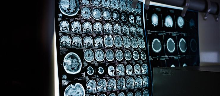
While planning an operation, surgeons must obtain the most precise image possible of the part of the body undergoing surgery. Researchers from the Department of Biomedical Engineering at the University and University Hospital of Basel have now developed a process that uses computed tomography data to transform two-dimensional images into three-dimensional virtual environments in real time.
Doctors can use the latest generation of virtual reality glasses to interact with, for example, the bone that requires surgery, viewing it from any desired angle and zoom distance. The sophisticated programming even means that fluid shadow rendering, which is important for creating an impression of depth, can be realistically calculated.
“Virtual reality offers the doctor a very intuitive way to obtain a visual overview and understand what is possible,” commented Philippe C. Cattin, research team leader, in a University of Basel statement.
Increasingly powerful hardware in the computer games industry supported the development of SpectoVive. The medical application has “helped rewire my senses,” according to Peter Maloca, who works at University Hospital Basel’s laboratory for optical coherence tomography (OTClab) and at Moorfields Eye Hospital in London.
He added: “Those who stand to gain the most here are doctors, their patients, and students – all of whom can share in this new information.” Some museums have also expressed interest in SpectoVive, seeing it as an opportunity to allow visitors to discover the world inside mummies or other exhibits.


